A Guide to Citrus Disease Identification
Citrus trees in both commercial and dooryard plantings can exhibit a host of symptoms reflecting various disorders that can impact their health, vigor, and productivity to varying degrees. Identifying symptoms correctly is an important aspect of management, as inappropriate remedial applications or actions can be costly and sometimes detrimental.
Disease symptoms addressed in this publication are an important aspect of commercial citrus production programs. Proper disease identification is an important factor in planning and conducting any disease control program. Disease symptoms may vary in expression on foliage, stems, roots, and fruit, and may not in all cases resemble those illustrated in various publications. Symptoms can vary considerably from mild to severe depending upon infection period, climatic conditions, and age of tissue when infection occurred. When in doubt about disease identification, seek advice before committing to costly and perhaps inappropriate corrective measures.
Greasy Spot (Zasmidium citri-griseum, formerly known as Mycosphaerella citri)
Infection by greasy spot produces a swelling on the lower leaf surface. A yellow mottle appears at the corresponding point on the upper leaf surface. The swollen tissue starts to collapse and turn brown and eventually the brown or black symptoms become clearly visible (Figure 1). Infection causes premature leaf drop, which occurs mostly in winter and early spring.
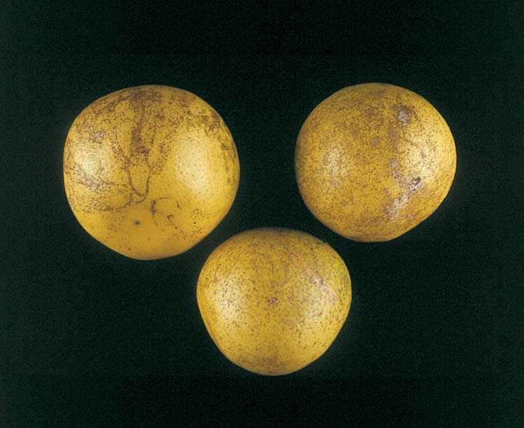
Greasy Spot Rind Blotch (Zasmidium citri-griseum, formerly known as Mycosphaerella citri)
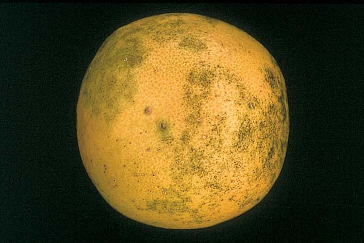
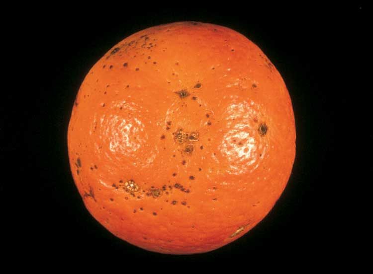
Pinpoint black specks occur between the oil glands with infection on grapefruit. When specks coalesce, they give rise to a symptom called pink pitting or greasy spot rind blotch (Figure 2). The living cells adjacent to the specks often retain a green color for much longer than normal or even when the fruit is degreened by ethylene.
Scab (Elsinoë fawcettii)
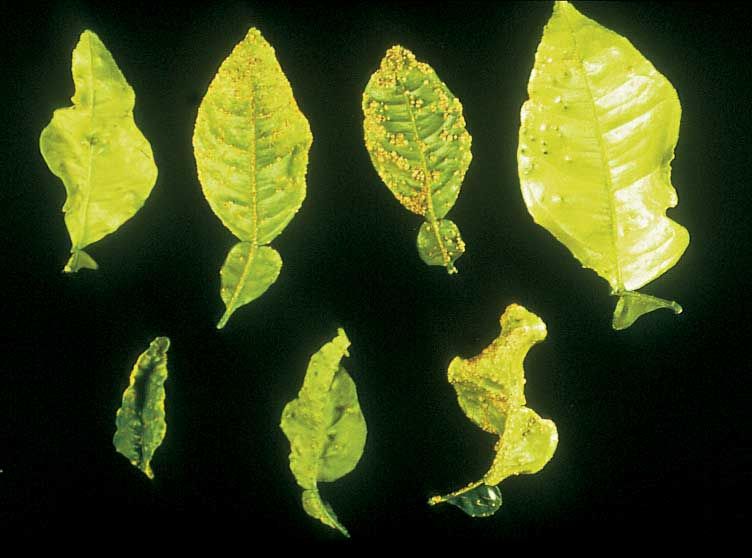
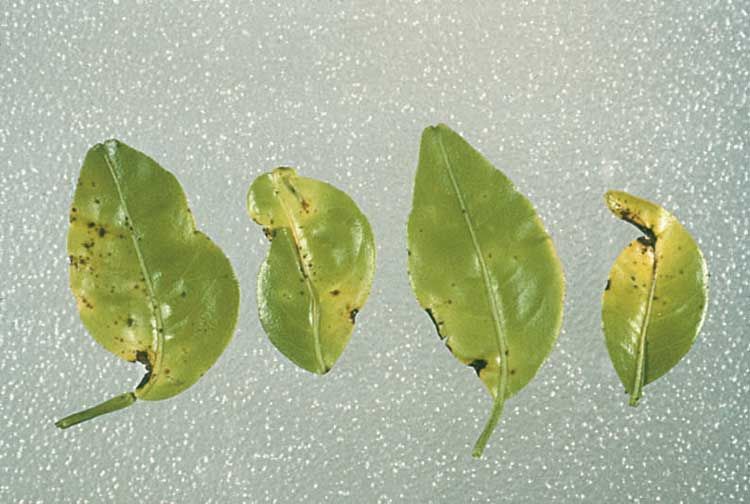
Small, pale orange, somewhat circular, elevated spots on leaves and fruit are the first evidence of the disease. As the leaves develop, the infection becomes well-defined, wart-like structures or protuberances on one side of the leaf, often with a conical depression on the opposite side (Figure 3). The crests of the wart-like growths usually become covered with a corky pale tissue and become somewhat flattened as the fruit matures, especially on grapefruit. The pustules may group together, covering large areas of the fruit surface (Figure 4) or leaves. Badly infected leaves become very crinkled, distorted, and stunted. Fruit severely attacked when very small often become misshapen.
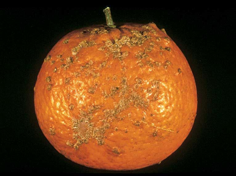
Scab can be particularly severe on Temples and lemons and often troublesome on Murcotts, Minneola tangelos, and grapefruit.
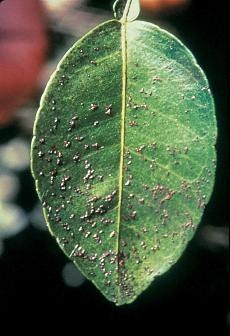
Melanose on Fruit (Diaporthe citri)
Lesions are small, raised, superficial dots, pustules, and irregularly shaped spots ranging from brick red to black (Figure 5). They feel like sandpaper when touched. Pustules are larger and more raised on grapefruit than on round oranges and tangerines.
The pustules often form in a tear-streaking pattern caused by water flowing down the surface and carrying spores. Fruit becomes resistant to infection about 12 weeks after petal fall.
Spores of Diaporthe citri arise from fruiting structures that develop on twigs that have recently died. The spores are moved to fruit via rain. Infected fruit does not serve as an inoculm source.
Phomopsis stem-end rot of mature fruit after harvest is also caused by Diaporthe citri.
Melanose on Leaves (Diaporthe citri)
On foliage, melanose first appears on the young leaves as minute, dark circular depressions with yellowish margins. Later they become raised, are rough, brown in color, and the yellow margins disappear (Figure 6). Leaves infected when very young may become distorted. Infested leaves do not serve as an inoculum source. Young green twigs can be infected.
In most cases, it is difficult and not economical to try to control melanose on the foliage.
Star Melanose
"Star melanose" occurs when copper is applied late during hot, dry weather and is due to copper damage to leaves. It has no relationship to melanose but may resemble symptoms of that disease. Copper causes the developing tissues to become more corky and darker than normal and the shape of the lesion often resembles a star (Figure 7).
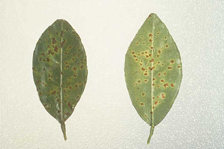
Alternaria Brown Spot (Alternaria alternata)
This fungus attacks fruit, leaves, and young shoots of susceptible varieties. Dancy tangerines and Minneola tangelos are the most severely affected varieties. Murcotts, Orlando tangelo, Lee, Nova, and Sunburst are often affected.
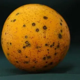
On fruit, the first symptoms appear as small, slightly depressed black spots that can cause the young fruit to fall from the tree (Figure 8). Fruit is usually immune to infection after reaching 3 to 4 months of age.
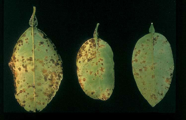
On susceptible varieties, only the young leaves and shoots can be infected. When infected they will produce brown, necrotic, blighted areas of various sizes, usually surrounded by yellow halos on the plant tissue (Figure 9). Infected foliage and fruit may prematurely fall from the tree with occasional shoot dieback.
Postbloom Fruit Drop (PFD) (Colletotrichum acutatum)
The disease appears as peach- to brown-colored necrotic spots on petals of flowers (Figure 10) and produces fruit drop and the formation of persistent buttons (calyces) which remain attached to stems.
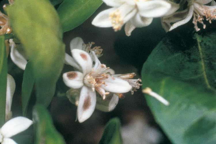
Damage is most severe on navel and Valencia oranges but can be found on all varieties of citrus including lemons and limes.
PFD is highly moisture dependent. Free water and high humidity are needed for infection, but windblown rain and/or rain splash are needed for dispersal of the spores from infected tissues to nearby healthy bloom.
The pathogen survives on the surface of leaves, twigs, and buttons between flowering periods.
Foot Rot (Phytophthora nicotianae or P. palmivora)
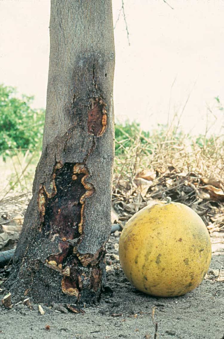
Lesions on a tree trunk usually occur on the bark at or just above the budunion on susceptible scions.
The lesions will first appear as a drop of gum on the surface of the bark. Upon further investigation, a brown, discolored, necrotic, slippery area will be found under the bark. In some cases, the margin of the infected area will break away from the healthy area and may curl back. Lesions can eventually girdle the entire tree trunk (Figure 11).
When the trunk is partially girdled by foot rot, the tree foliage may become dull and chlorotic, with the mid and main lateral veins of each leaf becoming yellow whereas the rest of the leaf remains nearly normal. This is similar to yellow vein chlorosis which is caused by nitrogen deficiency.
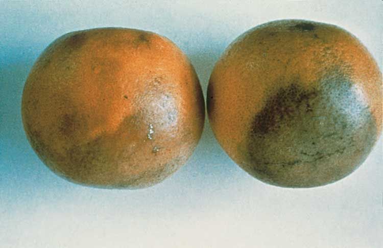
Brown Rot of Fruit (Phytophthora species)
Infected fruit exhibit a light brown, leathery decay which is not sunken below adjacent rind and produces a characteristic pungent, rancid odor (Figure 12). Under humid conditions, white mycelium is produced on rind surface. Fruit may become infected via soil contact, splash dispersal with soil particles, or fruit-to-fruit spread by windblown rain. Infected fruit prematurely fall from the tree. All cultivars may be affected with early season varieties showing greater susceptibility.
Citrus Canker (Xanthomonas citri)
Raised rough lesions are produced on young fruit and leaves of citrus (Figure 13). Bacteria are produced under moist conditions and dispersed by windblown rains. Bacteria enter leaf stomates or wounds on leaves, twigs, or fruit.
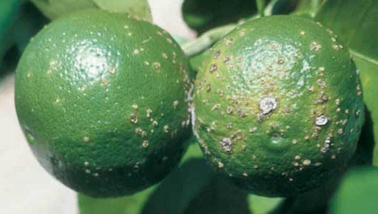
Citrus canker reduces yields, fresh fruit quality, and increases leaf defoliation.
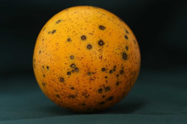
Citrus Black Spot (Phyllosticta citricarpa formerly known as Guignardia citricarpa (sexual stage), Phyllosticta citricarpa (asexual stage))
Black spot is found on all commercial citrus species, except Tahiti lime, in many subtropical regions with high summer rainfall. The causal agent is Guignardia citricarpa (sexual stage), Phyllosticta citricarpa (asexual stage), which reproduces on fallen, infected leaves.
Infected fruit is not suitable for fresh market but can be processed. Black spot can cause fruit drop and reduce yields. Symptoms on fruit or leaves appear as small, round, sunken, necrotic spots with gray centers and may be surrounded by green tissue or dark brown ring and yellow halo.
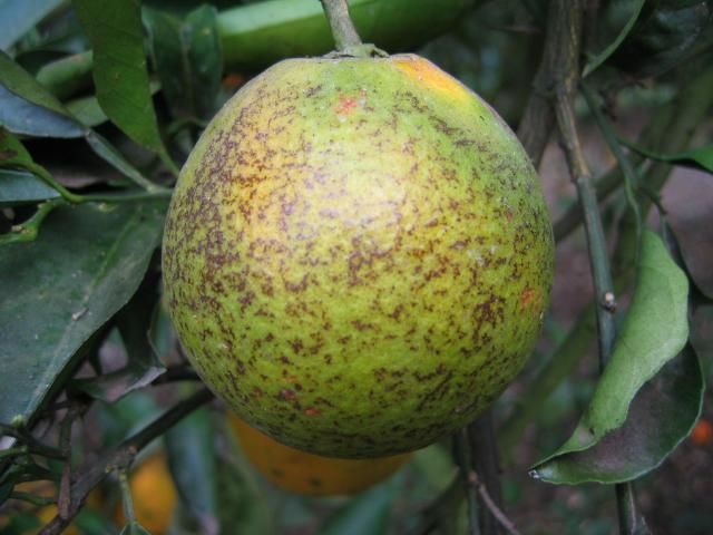
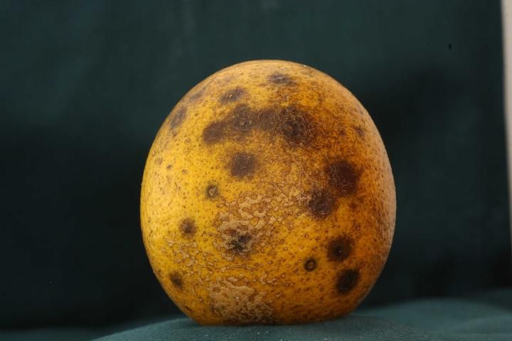
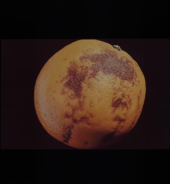
Black spot symptoms vary widely and have different names to best describe the symptoms. Hard spot symptoms (Figure 14) are about 0.1 to 0.4 inch in diameter and are nearly circular depresses with gray necrotic tissue in the middle of the spot and with brick-red to black margins that may be cracked around the edges. False melanose symptoms (Figure 15) appear on green fruit early in the season. The lesions are 0.04 to 0.12 inch in diameter and vary in color from tan to chocolate brown. False melanose symptoms can develop into hard spot as the season progresses. Cracked spot (Figure 16) is reported to be an interaction between rust mites and black spot fungus. Cracked spots are large, diffuse, smooth lesions that form raised cracks. Virulent spot (Figure 17) start as an irregularly shaped, sunken lesion with a reddish color. When the spots group together, they turn brown to black and the older lesions can become leathery. Virulent spot is found more on mature, older fruit at the end of the season.
Credit: Megan M. Dewdney, UF/IFAS CREC
Credit: Megan M. Dewdney, UF/IFAS CREC
Credit: Megan M. Dewdney, UF/IFAS CREC
Credit: Megan M. Dewdney, UF/IFAS CREC
Fruit remain susceptible for 4–5 months after fruit set. Disease pressure is greater on older trees than on younger ones.
Fungicide applications must be carefully timed to coincide with infection periods. Frequent sprays (up to 5) during the susceptible period may be required in heavily infested blocks.
More information on citrus disease identification and control is available at https://crec.ifas.ufl.edu/resources/production-guide#diseases.